Any area that looks abnormal. ABNORMAL CHEST XRAY 2.
 The Radiology Assistant Basic Interpretation
Today well talk about additional interesting chest x-ray findings including nodules masses atelectasis scarring pneumothorax pleural effusion rib fractures and heart failure.
Abnormal chest x ray. When viewed against a lit background your doctor can look for an array of problems from tumors to broken bones. It could be one problem or a litany of problems or it could be nothing serious. An abnormal chest scan could mean many things.
If you are told that your X-Ray is abnormal it can mean a few things. If there is an area that is different from the surrounding ipsilateral lung then this is likely to be the abnormal area. The condition of your lungs.
The chest contains our airways or wind pipes which are the hollow tube that act as a passage for the air to flow into our lungs during breathing lungs themselves blood vessels of the lungs and the space between the outer lining of the lungs and the chest wall. The bronchoscope is then passed gently through your nose to the back of your throat and down into your lungs. Chest X-rays can detect cancer infection or air collecting in the space around a lung which can cause the lung to collapse.
Lung Parenchyma Pleura Hilum Mediastinum Diaphragm Chest wall and bones 3. A chest x-ray is a radiology test that involves exposing the chest briefly to radiation to produce an image of the chest and the internal organs of the chest. Chest for a review of basic chest anatomy.
In this way doctors can examine the heart lungs bones and blood vessels. Nodules Masses and Tumors Quick summary of terminology. Normal chest anatomy normal chest x-rays abnormal chest x-rays part I.
This image shows a normal chest. What Abnormal Results Mean. In some cases a chest X-ray can detect a pulmonary nodule a small spot on your lung.
Chest radiographic findings of ABPA include lobar infiltrates 1314 perihilar glovelike tubular shadows representing mucus-filled bronchiectasis 1314 and tram-line bronchial walls due to. Abnormal lung An attempt to get the whole book in one small presentation. National Library of Medicine.
Introduction This presentation is just an effort to classify the visualized abnormalities by their radiological appearances. A chest x-ray is an x-ray of the chest lungs heart large arteries ribs and diaphragm. Standard x-rays are performed with the patient standing facing an x-ray film or digital cassette 6 feet away from an x-ray tube.
A normal chest x-ray can be used to define and interpret abnormalities of the lungs such as excessive fluid pneumonia bronchitis asthma cysts and cancers. Whilst sitting in an upright position the inside of your nose will be lubricated with a gel and your throat sprayed with a local anaesthetic this may taste rather bitter. They can also show chronic lung conditions such as emphysema or cystic fibrosis as well as complications related to these conditions.
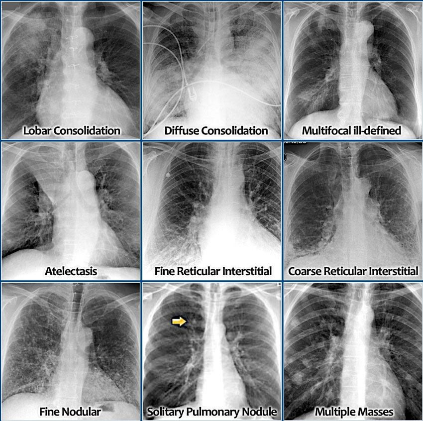
Abnormal results may be due to many things including. Homogeneous shadows grouped according to shape size and distribution. If the X-ray images show abnormalities this means that there is.
Chest X-ray examination is a mandatory part of visa medical examinationThere can be an abnormality on chest x ray which may lead to rejection on visaMost c. Parenchymal diseases Increased radiographic density Predominantly Airspace Predominantly Interstitial tissueDecreased radiographic density 4. Check out Anatomy for Radiology.
But in order to understand what these abnormal findings look like on a chest x-ray its important to first learn what a healthy chest x-ray looks like. Some of the common reasons to order a chest X-ray test are cough shortness of breath chest pain poor oxygenation hypoxia back pain chest injury and fever. A nodule is smaller than 3 cm in diameter a mass.
Rolando Sanchez MD says an abnormal chest x-ray could show an enlarged heart fluid in the lungs air pockets pneumonia among many other things. Certain abnormalities detected on the doctors physical examination of the lung heart or chest wall abnormal heart sounds abnormal lung sounds chest wall deformity etc. What is an abnormal chest x-ray.
A lab usually develops the images from a chest X-ray on large sheets of film. Abnormal Chest xray 1. The chest x-ray is one of the most common imaging tests performed in clinical practice typically for cough shortness of breath chest pain chest wall trauma and assessment for occult disease.
A chest X-ray is an imaging test that utilises low doses of radiation in short blasts to create images of the inside of a patients chest. Once you have spotted asymmetry the next step is to decide which side is abnormal. An abnormal chest X-ray isnt always a cause for concern but it signals a need to gather more information.
Your doctor may want to take a tissue sample biopsy of the nodule to look for signs of cancer but less than five percent of these nodules are cancerous. Abnormal chest xray ppt 1. Asymmetry of lung density is represented as either abnormal whiteness increased density or abnormal blackness decreased density.