This type of core needle biopsy is done under guidance of an MRI an imaging technique that captures multiple cross-sectional images of your breast and combines them using a computer to generate detailed 3-D pictures. During your biopsy an imaging physician will use an MRI scanner to accurately guide a needle to the biopsy.
 Mri Guided Breast Biopsy A Review Of Technique Indications And Radiological Pathological Correlations Sciencedirect
Visit our website or contact us on 1-888-360-0001.
Mri guided breast biopsy. Outcome of MRI-guided vacuum-assisted breast biopsy initial experience at Institute of Oncology Ljubljana Slovenia Marta Zebic-Sinkovec K. Once the area to biopsy is found your radiologist will guide a thin needle into your breast. A biopsy can help diagnose abnormalities such as infection inflammation or malignancy.
All facilities that perform breast MRI should have the ability to perform MRI-guided biopsy. MRI Breast biopsy is a method of performing a biopsy of an area of concern that has been best imaged using Magnetic Resonance Imaging MRITo learn more pl. MRI Guided Breast Biopsy.
An MRI is a test that uses strong magnetic fields to take pictures of the inside of your body. Board-certified radiologists will then diagnose these cells to determine what further treatment is required. Your doctor has requested an MRI-guided breast needle-core biopsy.
MR biopsy technique Prone positions Open-configuration Breast Coil Use Grid compression device to aid localization MR compatible biopsy device multiple commercial options. The ability to perform MR-guided breast biopsy or localization therefore becomes an integral component of a dedicated breast MRI program. MR biopsy capability is required component a breast MR imaging practice.
Image Guided Biopsy In many cases an imaging technique such as an ultrasound or MRI is used in combination with a biopsy to provide the most accurate reading possible. Sometimes these abnormalities turn out not to be a problem. The goal of a biopsy is to remove a small sample of tissue for testing in a laboratory.
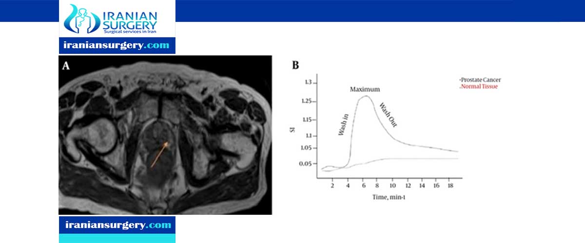
The patients were scanned using a 15-tesla or 3-tesla whole-body MRI scanner equipped with a breast coil. Magnetic Resonance MRI-Guided Breast Biopsy. An MRI breast biopsy is a procedure that uses computer technology to guide a needle to an abnormality seen on MRI.
Do not take anti-inflammatory products 7 days before the biopsy. The cancer rate of patients who undergo MRI-guided breast biopsy has been reported to range from 20 to 61 depending on the study design and population 614. MRI-guided core needle biopsy.
Early on MRI biopsies were performed by placing either a wire or a titanium clip with MRI guidance into the area of enhancement to guide surgical excision. During this procedure you lie facedown on a padded scanning table. Although it is predictable that this variability comes from differences between cohort groups with certain characteristics to our knowledge no detailed study has compared the cancer rate with the characteristics of such groups and estimated the biopsy yield in a general practice population.
Hertl Maksimiljan Kadivec Mihael Cavlek Gasper Podobnik M. Using MRI guidance and a vacuum-assisted biopsy system the authors removed seven to 12 tissue samples from each patient and marked the biopsy incision site at the tumor location before the start of chemotherapy. According to the American College of Radiology ACR practice guidelines MRI-guided breast biopsy is warranted for suspicious lesions or lesions highly suggestive of malignancy BI-RADS 4 and 5 that are mammographically and sonographically occult or seen with certainty only on breast MRI.
An MRI-guided breast biopsy is a non-radiation minimally invasive technique used to gather tissue samples from a breast abnormality. A breast biopsy is done to take samples of tissue from your breast to examine it for cancer. If your physician refers you for an MRI-guided breast biopsy physicians will use MRI imaging to guide a hollow needle into the breast to remove cells from the area of concern.
Magnetic resonance- or MR-guided breast biopsy uses a powerful magnetic field radio waves and a computer to help locate a breast lump or abnormality and guide a needle to remove a tissue sample for examination under a microscope. When a suspicious finding is seen on a breast magnetic resonance imaging MRI examination and other imaging and clinical examinations are negative the only way to perform a biopsy of the lesion is by using MRI guidance. Your radiologist doctor who specializes in image-guided procedures has recommended that you have an MRI-guided breast biopsy.
WHAT IS AN MRI Magnetic Resonance Imaging-GUIDED BIOPSY. It does not use ionizing radiation and leaves little to no scarring. Do not take blood thinning products 7 days before the biopsy.
Non-mass enhancement at breast MRI is defined in the BI-RADS lexicon as an area of enhancement that does not meet criteria for a mass such as by having nonconvex borders or intervening fat or fibroglandular tissue between the enhancing component. If there is a potential problem early detection is essential and increases treatment options and the likelihood of successful recovery. Do not take aspirin or aspirin products 7 days before the biopsy.
You will first have an MRI done to find the exact area of your breast to biopsy. The abnormality usually cannot be felt on breast self-examination or clinical examination by your primary care physician or seen by mammogram or ultrasound of the breast. Bonnie Joe describes how a breast MR-guided biopsy is performed.
Guided breast biopsy ultrasound from Cura4U and get up to 80 discount on all radiology services.
How painful is a biopsy of the prostate. The prostate is a small walnut-shaped gland in men that produces fluid that nourishes and transports sperm.
 Prostate Needle Biopsy Recovery Prostate Biopsy Recovery Time
Samadis prostate surgery takes just 15-2 hours and almost all of his patients return home the day after having a robotic prostatectomy.

Prostate biopsy recovery period. A prostate biopsy is a when a doctor removes small samples of tissue from your prostate to test for cancerYour doctor will order one if the results from a screening a blood test or a digital. A prostate biopsy can often determine if there is cancer present. This procedure typically takes about 15 minutes to perform.
Miller recommends a minimum waiting period of 8 weeks following biopsy prior to proceeding with robotic prostatectomy. After this biopsy I could not urinate. Over the years he continually told me I had a large but smooth prostate.
A prostate biopsy is a procedure to remove samples of suspicious tissue from the prostate. One went to a movie and the other went out. It may continue for up to three days.
How soon can I exercise after a prostate biopsy. Prostate Biopsy Recovery Period. Burning with urination It is normal to feel burning with urination for the first 24 hours after the biopsy.
Some people may notice a rust- or red-colored tint to their semen for several weeks after their biopsies notes Mayo. Prostate biopsy results What percentage of prostate biopsies are positive for cancer. Significant inflammation occurs after a prostate biopsy resulting in temporary distortion of the anatomy particularly when operating under such extreme magnification.
The recovery time following a prostate biopsy is several days according to Mayo Clinic. The biopsy needle will be removed and firm pressure will be applied to the biopsy site until the bleeding has stopped. Instructions Following Prostate Biopsy What can I expect after a prostate biopsy.
One report indicated that various studies showed that between 5 of men and 90 of men had blood in their semen after a biopsy. So its nothing to worry about. A few months ago I had my third biopsy over a 10 year period.
Most people feel slight soreness or experience some light rectal bleeding or blood in the stool or urine for a few days following the procedure. During a prostate biopsy a needle is used to collect a number of tissue samples from your prostate gland. Jennifer Whitlock RN MSN FNP-C is a board-certified family nurse practitioner.
A doctor may recommend a prostate biopsy if you have an elevated prostate-specific antigen PSA test or abnormal digital rectal exam which can indicate prostate cancerWhile screening tests may suggest there is a problem a prostate biopsy is needed to make a prostate cancer diagnosis and determine the aggressiveness of the disease. After the biopsy it is normal to experience the following sensations or symptoms. Is This an Emergency.
Some discomfort is likely during the recovery time but sometimes more severe complications can arise. It has survived not only five centuries but also the leap into electronic typesetting. Can you drive after a prostate biopsy.
There are a few symptoms that the patient may experience for several days to weeks after the procedure. The prostate is part of the male reproductive system and sits underneath the bladder surrounding the urethra which is the urine outflow tube. Both swear absolutely swear that it was a walk in the park.
The biopsy needle will be inserted through the incision and into the prostate several times to get samples from different parts of the gland. She has experience in primary care and hospital medicine. A similar study had very similar results.
The gland grows or enlarges with age. Both worked the next day. One thing that lasts for a long time is blood in semen.
Recovery after prostate surgery has several elements from basic needs like taking care of your wound and managing issues like constipation to more involved. More amazing to me is that they did activities the day of the biopsy. Nly five centuries but also the leap into electronic typesetting industry.
Men that do prostate biopsy usually notice blood in their semen for up to 6 weeks. My father died of PC so as my psa rose to 97 and my urologist had tried all the usual tests PCA etc it was time for the biopsy again. But usually it resolves after 2 days of doing the biopsy.
Lorem Ipsum is simply dummy text ever since the 1500s when an unknown printer took a galley of type and scrambled it to make a type specimen book. When eliminating men who did not ejaculate after the procedure they found about 90 had blood in the semen that lasted around 4 weeks and 6 ejaculations after the procedure. If you are referring to bleeding assuming you are not on any blood thinning medications it could be anywhere up to around 2 weeks.
Following proper prostatectomy recovery guidelines patients who experienced normal continence prior to surgery should regain function within 12-13 months. Casey Ng answered 17 years experience Urology bleeding. Overall I hated my biopsy more than my robotic surgery.
I have two friends that had the exact type of biopsy one prior to mine one after. How long does it take to recover prostate biopsy.
Sometimes a local anaesthetic is used. In a biopsy your surgeon will take a small section of tumor and send it to a pathologist who will look at it under a microscope to make or confirm a brain tumor diagnosis.
 Brain Biopsy Of A Suspected Cerebellar Lymphoma Journal Of Medical Insight
Pediatric oncologists use the data collected from sequencing both the tumor and the young patients healthy tissue to develop personalized treatment recommendations.

Brain tumor biopsy procedure. Magnetic resonance imaging have been insufficient in showing the cause of symptoms and if it is felt that the benefits of histological diagnosis will influence the treatment plan. Your healthcare provider will talk to you about how to prepare for your procedure. A special frame may be put on the head to hold it in place.
Given the potential risks surrounding the procedure cerebral biopsy is indicated only if other diagnostic approaches eg. The sample can be tested for cancer infection or brain disease. He or she will insert a needle through the hole and into your brain.
A biopsy can be performed as a separate procedure if. You usually have a biopsy of your brain under general anaesthetic. Performed in the operating room the procedure involves the removal of a small piece of tissue most commonly from the brain but could include samples from the scalp blood vessels or dura mater the outermost membrane covering the brain.
The biopsy will provide information about the type of tumor. How to prepare for a brain biopsy. A brain biopsy is a procedure to remove a sample of tissue from your brain or tumor.
Stereotactic biopsy is a minimally invasive procedure. It is usually performed at the same time as the surgery to remove a brain tumor called an open biopsy. The biopsy is guided by computed tomography CT or magnetic resonance imaging MRI scans.
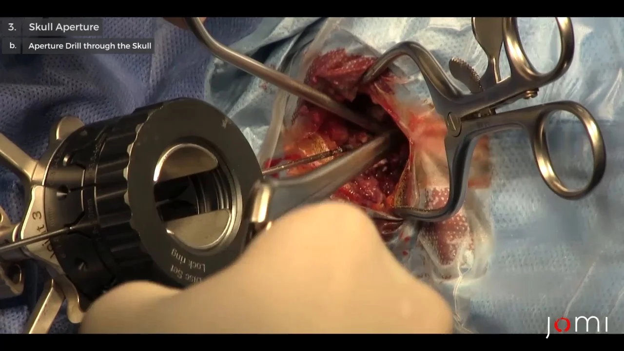
Your neurosurgeon drills a small hole into your skull. A Stereotactic Brain Tumor Biopsy is a neurosurgical procedure in which samples of tissue are taken from the tumor site. If the person has a brain tumor biopsy is 95 sensitive.
Stereotactic biopsy procedures in which a computer-based three-dimensional-image-guided system accurately locates patients brain tumors are relatively new diagnostic methods. Your incision will be closed with stitches or staples. A brain biopsy may be conducted to determine whether a tumor found in the brain is cancerous or benign.
Stereotactic Brain Biopsy is a common procedure that allows a neurosurgeon to diagnose a brain lesion. This helps your doctors decide the best treatment for you. Because a biopsy is no less invasive than brain surgery your doctors will try to remove the whole tumor during a biopsy procedure so that you will not need another surgery if possible.
A stereotactic biopsy uses 3-D imaging technology as well as data from CT and MRI scans to examine a tumor or a piece of the brain. Biopsy for brain and spinal cord tumours. You usually have a biopsy taken under a general anaesthetic.
The procedure is usually done under general anesthesia. Stereotactic biopsy is often used if a tumor is in a part of the brain that is hard to reach or near a vital area. More often it is done as part of a larger operation to remove the tumour.
Sometimes this is done before you have any other treatment. Surgery can also provide an opportunity to biopsy the tumor to learn more about what type it is and whether its cancerous. Resection is the surgical removal of all or part of the tumor itself.
The tumor cannot be removed without damaging critical parts of the brain. Prior to the biopsy a CT scan is done to locate the specific area of the brain where the sample will be taken from. Technical aspects of the procedure should undoubtedly reflect on its success rate and.
A bandage will be placed over your incision. A select group of pediatric neurosurgeons are using intraoperative MRI to guide them through the precarious procedure of excising brain tissue for biopsy. What will happen after a brain biopsy.
MRI or CT pictures may be taken to help your provider find the tumor. Typically patients present with symptoms that require a physician to capture images of the brain. A brain tumor biopsy requires a surgical procedure under general anesthesia usually involving removal of a section of the skull to access the brain tissue.
A biopsy can be performed as part of an operation to remove the brain tumor or a biopsy can be performed using a needle. A biopsy is when the surgeon removes a piece of the brain tumour. Generally speaking there are two reasons for brain tumor surgery.
BackgroundEndoscopic biopsy of brain tumors is an important part of the armamentarium of management of intra- and periventricular tumors that is generally considered an acceptable and in some situations a preferred method for tissue samplingThe diagnostic yield of the procedure has been variably reported. A biopsy means taking a small tissue sample from your brain and looking at it under a microscope. Complications from stereotactic biopsy procedures are minimal compared with open craniotomy procedures because they are performed with local anesthesia.
A brain biopsy is a procedure to remove a sample of abnormal tissue for examination under a microscope. The goal of brain tumor surgery is to remove as much of the tumor as possible without causing harm to normal tissue. The purpose of a biopsy is to discover the type and grade of a tumor as well as its molecular biology and its growth pattern.
The provider will remove pieces of the tumor with the needle. The tissue cells taken during the biopsy can show what kind of brain lesion abscess tumor is present and whether it is benign not cancerous or cancerous malignant. The first treatment for a brain tumor is often surgery.
What you need to know about a brain biopsy. A biopsy is a surgical procedure to remove a small sample of brain tumor tissue for examination under a microscope. This means that you will be asleep and wont feel anything.
A stereotactic needle biopsy may be done for brain tumors in hard to reach areas or very sensitive areas within your brain that might be damaged by a more extensive operation.