The general examination for chest is PA posterior anterior and the lateral LAT chest X-ray. How Much Does a Chest X-ray Cost.
 How Much Does Chest X Ray Cost For Stroke Strokebelt Org
This price includes the doctors order that is required to get a chest x-ray all x-ray fees and a copy of your results that includes a radiologist interpretation.
Cost chest x ray. You will be able to go soon after the chest X-ray is finished and can continue with normal activities. As with all X-rays there is a small exposure to radiation. Check Chest X-ray cost near you in DelhiNCR.
It helps spot any abnormalities in the chest. The standard chest examination consists of a PA posterioranterior and lateral chest x-rayThe films are read together. The procedure itself is relatively inexpensive minimally invasive and painless for your pet but it requires the animal to remain still for an extended period of time.
On MDsave the cost of an X-ray ranges from 60 to 320. CXR test produces pictures of the Chest. A normal chest x-ray can be used to define and interpret abnormalities of the lungs such as excessive fluid pneumonia bronchitis asthma cysts and cancers.
It also helps in detecting the accumulation of fluids in lungs or gut. The PA exam is done in the view of the patient as if standing with their right side towards your left side. Chest x-rays CXR are a scan used to evaluate the lungs heart and chest wall and can detect medical conditions such as.
On MDsave the cost of a Chest X-ray ranges from 54 to 438. A chest x-ray is a radiology test that involves exposing the chest briefly to radiation to produce an image of the chest and the internal organs of the chest. Costs of chest X-rays.
The typical price of chest x-ray is in between 200 400 dollars. A chest X-ray is the most commonly performed procedure in any X-ray department and can be performed in the department as an outpatient or on the wards with a mobile X-ray machine. The PA exam is viewed as if the patient is standing in front of you with their right side on your left.
A chest X-ray gives a black-and-white picture of your lungs ribs heart and diaphragm. Alert Your chest x-ray and medical examination results must be no more than 3 months old when we receive your application. Actual X-ray cost depends on the provider the part of the body being X-rayed and the number of views taken.
Those on high deductible health plans or without insurance can shop compare prices and save. It could also be more. There are also x-ray packages with the cost that depends on the provider and the number of views taken.
Get the best price for a Chest X-ray here and other X-rays with 50 discount. Mammogram helps in looking for breast cancer. Submit a request for further information a quotation or indicative cost.
So here it is. For uninsured clients on average a chest X-ray will cost around 420 with a range from 120 to 2100. Both films are read together for a proper analysis.
Cost depends on the provider and the number of views taken. X-rays are mainly used for examining broken bones but there are several other uses. After the chest X-ray.
X-ray chest Bone Densitometry Doppler Studies ECG Echo EEG EMG Extra Film CD Mammography TMT UltraSound UROFLOWMETRY X-RAY NCV Others PFT SSEP X-RAY FACE NECK X-RAY SKULL X-RAY SHOULDER HANDS Special Investigation Color X-RAY X-RAY SPINE X-RAY ABDOMEN X-RAY CHEST X-RAY HIP KNEE LEG ANKLE FOOT X-RAY STERNUM. Public patient no cost to you unless advised otherwise. For an Australian patient in a public hospital in Western Australia.
If you need to get a chest x-ray or medical examination make sure you have all your visa application documents ready to submit to us once your x-ray or medical examination is completed. The typical cost of X-rays in dogs is about 150 to 250. We have provided an indication of the cost of a private chest x-ray based on the prices published by several of the major providers of private surgery.
How Much Does an X-ray Cost. The average cost however is 370. Those on high deductible health plans or without insurance can shop compare prices and save.
Your enquiry will be forwarded to up to 3 private healthcare providers. Average Cost of Chest X-ray Well so how much does a chest x-ray cost. A chest X-ray can be useful in diagnosing pneumonia.
These prices include a frontal frontal and lateral or complete chest x-ray. This is much more difficult to accomplish with a dog than with a human. X-ray also helps in detecting heart issues by examining the size and shape of the heart.
The total fee for a screening chest x-ray through Accesa Health is 95.
Any area that looks abnormal. ABNORMAL CHEST XRAY 2.
 The Radiology Assistant Basic Interpretation
Today well talk about additional interesting chest x-ray findings including nodules masses atelectasis scarring pneumothorax pleural effusion rib fractures and heart failure.
Abnormal chest x ray. When viewed against a lit background your doctor can look for an array of problems from tumors to broken bones. It could be one problem or a litany of problems or it could be nothing serious. An abnormal chest scan could mean many things.
If you are told that your X-Ray is abnormal it can mean a few things. If there is an area that is different from the surrounding ipsilateral lung then this is likely to be the abnormal area. The condition of your lungs.
The chest contains our airways or wind pipes which are the hollow tube that act as a passage for the air to flow into our lungs during breathing lungs themselves blood vessels of the lungs and the space between the outer lining of the lungs and the chest wall. The bronchoscope is then passed gently through your nose to the back of your throat and down into your lungs. Chest X-rays can detect cancer infection or air collecting in the space around a lung which can cause the lung to collapse.
Lung Parenchyma Pleura Hilum Mediastinum Diaphragm Chest wall and bones 3. A chest x-ray is a radiology test that involves exposing the chest briefly to radiation to produce an image of the chest and the internal organs of the chest. Chest for a review of basic chest anatomy.
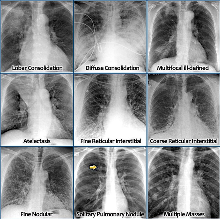
In this way doctors can examine the heart lungs bones and blood vessels. Nodules Masses and Tumors Quick summary of terminology. Normal chest anatomy normal chest x-rays abnormal chest x-rays part I.
This image shows a normal chest. What Abnormal Results Mean. In some cases a chest X-ray can detect a pulmonary nodule a small spot on your lung.
Chest radiographic findings of ABPA include lobar infiltrates 1314 perihilar glovelike tubular shadows representing mucus-filled bronchiectasis 1314 and tram-line bronchial walls due to. Abnormal lung An attempt to get the whole book in one small presentation. National Library of Medicine.
Introduction This presentation is just an effort to classify the visualized abnormalities by their radiological appearances. A chest x-ray is an x-ray of the chest lungs heart large arteries ribs and diaphragm. Standard x-rays are performed with the patient standing facing an x-ray film or digital cassette 6 feet away from an x-ray tube.
A normal chest x-ray can be used to define and interpret abnormalities of the lungs such as excessive fluid pneumonia bronchitis asthma cysts and cancers. Whilst sitting in an upright position the inside of your nose will be lubricated with a gel and your throat sprayed with a local anaesthetic this may taste rather bitter. They can also show chronic lung conditions such as emphysema or cystic fibrosis as well as complications related to these conditions.
Abnormal results may be due to many things including. Homogeneous shadows grouped according to shape size and distribution. If the X-ray images show abnormalities this means that there is.
Chest X-ray examination is a mandatory part of visa medical examinationThere can be an abnormality on chest x ray which may lead to rejection on visaMost c. Parenchymal diseases Increased radiographic density Predominantly Airspace Predominantly Interstitial tissueDecreased radiographic density 4. Check out Anatomy for Radiology.
But in order to understand what these abnormal findings look like on a chest x-ray its important to first learn what a healthy chest x-ray looks like. Some of the common reasons to order a chest X-ray test are cough shortness of breath chest pain poor oxygenation hypoxia back pain chest injury and fever. A nodule is smaller than 3 cm in diameter a mass.
Rolando Sanchez MD says an abnormal chest x-ray could show an enlarged heart fluid in the lungs air pockets pneumonia among many other things. Certain abnormalities detected on the doctors physical examination of the lung heart or chest wall abnormal heart sounds abnormal lung sounds chest wall deformity etc. What is an abnormal chest x-ray.
A lab usually develops the images from a chest X-ray on large sheets of film. Abnormal Chest xray 1. The chest x-ray is one of the most common imaging tests performed in clinical practice typically for cough shortness of breath chest pain chest wall trauma and assessment for occult disease.
A chest X-ray is an imaging test that utilises low doses of radiation in short blasts to create images of the inside of a patients chest. Once you have spotted asymmetry the next step is to decide which side is abnormal. An abnormal chest X-ray isnt always a cause for concern but it signals a need to gather more information.
Your doctor may want to take a tissue sample biopsy of the nodule to look for signs of cancer but less than five percent of these nodules are cancerous. Abnormal chest xray ppt 1. Asymmetry of lung density is represented as either abnormal whiteness increased density or abnormal blackness decreased density.
The WHO has released a consolidated summary of WHO recommendations on the use chest radiography in TB detection and guidance on programmatic approaches. A chest x-ray is a radiology test that involves exposing the chest briefly to radiation to produce an image of the chest and the internal organs of the chest.
 Pulmonary Tuberculosis Radiology Case Radiopaedia Org
Health care personnel with a positive TB test result should receive a symptom evaluation and a chest x-ray to rule out TB disease.

Tb chest x ray. The fourth component of a complete TB medical evaluation is a chest x- ray. A normal chest x-ray can be used to define and interpret abnormalities of the lungs such as excessive fluid pneumonia bronchitis asthma cysts and cancers. The CDC states that a positive test for TB infection only tells that a person has been infected with TB germs.
Additional workup may be needed based on those results. There are no radiological features which are in themselves diagnostic of primary mycobacterium tuberculosis infection TB but a chest X-ray may provide some clues to the diagnosis. In active pulmonary TB infiltrates or consolidations andor cavities are often seen in the upper lungs with or without mediastinal or hilar lymphadenopathy.
Each individual case is different and so questions on your follow-up cannot always be answered until the x-ray is reviewed. In our current release there are 3500 TB images and 3500 normal images. The contagious disease of tuberculosis not only attacks the lungs but also can affect other organs such as the kidney spine and brain.
Hover onoff image to showhide findings. Pulmonary manifestations of tuberculosis are varied and depend in part whether the infection is primary or post-primary. A posterior-anterior PA chest X-ray is the standard view used.
The chest X-ray represents the final step in tuberculosis TB testingPrior to receiving an X-ray an individual must test positive for TB via a PPD skin test or QuantiFERON QFT test. This scholarly work is published in IEEE Access Link. There are multiple light areas opacities of varying size that run together coalesce.
An X-ray is an imaging test that uses small amounts of radiation to produce pictures of the organs tissues and bones of the body. Chest radiography is an essential tool for the early detection of TB and therefore fundamental to achieve the targets set out in WHOs End TB Strategy. However TB may mimic other diseases on x-rays and non TB conditions may look like TB.
It may show infiltrates or cavities. Other views lateral or lordotic or CT scans may be necessary. A tuberculosis chest x-ray is a diagnostic procedure used to detect the presence of tuberculosis in the lungs.
The chest X-ray and classification worksheet by the Centers for Disease Control and Prevention CDC of the United States is designed to group findings into categories based on their likelihood of being related to TB or non-TB conditions needing medical follow-up. The mobile x-ray vans which will be moving round the State to provide prompt chest x-ray services and effective diagnosis of TB within the communities were manufactured to specification and are adequately equipped to effectively provide comprehensive on-site screening diagnosis and treatment for TB. Tuberculosis TB Chest X-ray Database A team of researchers from Qatar University Doha Qatar and the University of Dhaka Bangladesh along with their collaborators from Malaysia in collaboration with medical doctors from Hamad Medical Corporation and Bangladesh have created a database of chest X-ray images for Tuberculosis TB positive cases along with Normal images.
Tap onoff image to showhide findings. Chest x-rays serve as an invaluable adjunct in the diagnosis and follow-up of TB. The chest x-ray will usually appear abnormal when a patient has TB disease in the lungs.
A general discussion of tuberculosis is found in the parent article. After your chest x-ray is reported on a decision is made as to whether you will require further x-rays check-ups or treatment. And a discussion of other.
It does not indicate whether they have progressed to TB disease. In HIV and other immunosuppressed persons any abnormality. A patient should have a chest x-ray if he or she has a positive IGRA or TST result or has signs and symptoms of TB disease.
Health care personnel with a documented history of a prior positive TB test should receive a baseline individual TB risk assessment and TB symptom screen upon hire ie preplacement. This procedure is used as a secondary screening method in patients who have had a positive skin test and in patients who are at high risk for tuberculosis infection but have not had a positive skin test. In certain cases a patient may need a tuberculosis chest X-ray to confirm this diagnosis.
What is a chest X-ray. Thus chest x-rays are neither specific nor sensitive and so remain a supplement to microbiological tests such as microscopy PCR and culture. Arrows indicate the location of cavities within these light areas.
The research team managed to classify TB and Normal Chest X-ray images with an accuracy of 983. However lesions may appear anywhere in the lungs. Please make sure you give credit to us while using the dataset code and trained models.
The lungs are the most common site of primary infection by tuberculosis and are a major source of spread of the disease and of individual morbidity and mortality. The x-ray on the left clearly shows that the opacities are located in the upper area of the lungs toward the back. These chest x-rays show advanced pulmonary tuberculosis.