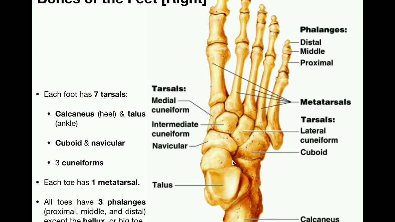
The foot is divided into three sections - the forefoot the midfoot and the hindfoot. This section of the foot is made up of five irregularly shaped bones called the tarsals.
 Anatomy Specific Bones Of The Feet Youtube
The extrinsic muscles arise from the anterior posterior and lateral compartments of the leg.

Anatomy of the foot. Some feet contain accessory ossicles or accessory bones Figure 9. In most two-footed and many four-footed animals the foot consists of all structures below the ankle joint. Some health conditions injuries and general wear and tear can all cause or contribute to conditions.
The fascia of the foot can be divided into superficial and deep fascia. The hindfoot forms the heel and. The foot contains 26 bones 33 joints and over 100 tendons muscles and ligaments.
Sechrest MD narrates an animated tutorial of the anatomy of the foot. The anatomy of the foot. In anatomy pronation is a rotational movement of the forearm at the radioulnar joint or foot at the subtalar and talocalcaneonavicular joints.
Heel arch digits and contained bones such as tarsals metatarsals and phalanges. The muscles acting on the foot can be divided into two distinct groups. Foot and ankle anatomy is quite complex.
The dynamic stability of the vault is achieved by the. In mammals that walk on their toes and in hoofed mammals it includes the terminal parts of one or more digits. They are mainly responsible for actions such as eversion inversion plantarflexion and dorsiflexion of the foot.
Common Ossicles of the Foot. Ebraheims educational animated video describes anatomical structures of the foot and ankle The Bony Anatomy The Joints Ligaments and the Compartment. The ankle and foot.
These all work together to bear weight allow movement and provide a stable base for us to stand and move on. It is much more fibrous in the sole and is thicker than in other areas of the foot making the heel act as a shock absorbing pad. Extrinsic and intrinsic muscles.
At the same time the foot must be strong to support more than 100000 pounds of pressure for every mile walked. In the foot there are two sesamoid bones located directly underneath the first metatarsal head embedded in the medial tibial side and lateral fibular aspect of the flexor hallucis brevis tendon. The foot is an extremely complex anatomic structure made up of 26 bones and 33 joints that must work together with 19 muscles and 107 ligaments to execute highly precise movements.
Bones of the human foot. These arches the medial arch lateral arch and fundamental longitudinal arch are created by the angles of the bones and strengthened by the tendons that connect the muscles and the ligaments. During the gait cycle the foot can pronate in many different ways based on rearfoot and forefoot function.
The ankle joint also known as the talocrural joint allows dorsiflexion and plantar flexion of the foot. The foot is responsible for balancing the bodys weight on two legs a feat which modern roboticists are still trying to replicate. This may sound like overkill for a flat structure that supports your weight but you may not realize how much work your foot does.
Find foot anatomy stock images in HD and millions of other royalty-free stock photos illustrations and vectors in the Shutterstock collection. The superficial fascia or the subcutaneous fat tissue is loose and deep to the dorsal skin. The midfoot is a pyramid-like collection of bones that form the arches of the feet.
These include the three cuneiform bones the cuboid bone and the navicular bone. Thousands of new high-quality pictures added every day. These extra bones are developmental variants.
This consists of five long metatarsal bones and five shorter bones that form the toes phalanges. Ligaments bind the bones to provide the static stability of the foot. The foot consists of thirty three bones twenty six joints and over a hundred muscles ligaments and tendons.
The arch of the foot plays a key role in weight-bearing and stability. Together they form the arch of the foot. The foot contains a lot of moving parts - 26 bones 33 joints and over 100 ligaments.
The anatomy of the foot is highly intricate consisting of many bones joints and ligaments. The last two together are called the lower ankle joint. The clinical names for these bones are the navicular cuboid and medial intermediate and lateral cuneiforms.
It is made up of three joints. The 26 bones of the foot create an architectural vault sup-ported by three arches and resting on the ground at three points which lie at the corners of an equilateral triangle Fig. Upper ankle joint tibiotarsal talocalcaneonavicular and subtalar joints.
Pronation of the foot refers to how the body distributes weight as it cycles through the gait. In this episode of eOrthopodTV orthopaedic surgeon Randale C.