Caffeine alcohol and over-the-counter OTC cold medications that contain pseudoephedrine can stimulate and irritate the hearts electrical conduction system and precipitate an episode of PSVT. SVT is caused by abnormal electrical impulses that start suddenly in the upper chambers of your heart the atria.
 Supraventricular Tachycardia Non Cardiac Surgery Cancer Therapy Advisor
What is supraventricular tachycardia SVT.

Paroxysmal supraventricular tachycardia causes. Wolff-Parkinson-White syndrome is a major risk factor for paroxysmal supraventricular tachycardia. The signal moves through the upper heart chambers the atria and tells the atria to contract. Apart from occasional detection by parents most tachycardias in this age group are revealed by heart failure signs such as poor feeding sweating and shortness of breath.
Causes and Risk Factors Paroxysmal supraventricular tachycardia may be set off by a premature heartbeat that rapidly signals the heart to be beat. There are four main types. It occurs when a short circuit rhythm develops in the upper chamber of the heart.
The cause is not known. Sometimes the signal messes up and keeps going around in a circle. Paroxysmal supraventricular tachycardia PSVT is a type of supraventricular tachycardia named for its intermittent episodes of abrupt onset and termination.
The contractions are caused by an electrical signal that begins in an area of the heart called the sinoatrial node also called the sinus node or SA node. Otherwise symptoms may include palpitations feeling lightheaded sweating shortness of breath and chest pain. PSVT is one of many.
PSVT can result because of underlying medical conditions or because of foods and medications that are ingested. In SVT electrical signals get caught in a loop that goes round and round. How can paroxysmal supraventricular tachycardia PSVT be prevented.
Supraventricular tachycardia SVT is an abnormally fast heart rhythm arising from improper electrical activity in the upper part of the heart. Supraventricular tachycardia SVT is a condition that causes your heart to beat much faster than it should. The pathways let electrical currents bypass the AV node which allows the currents to move too quickly to the ventricles.
In SVT the looping signals are above the ventricles. This results in a regular but rapid heartbeat that starts and stops abruptly. SVT is a type of abnormal heart rhythm called an arrhythmia that starts in the upper part of your heart.
It can then slow down abruptly. A muscle in the upper chambers of your heart sends out an electrical signal. Sometimes a problem with your heart s electrical signals can make it speed up even when youre not anxious or exercising.
But with SVT your heart rate suddenly goes above 100bpm. This may be caused by several abnormalities such as having. It may last from a few seconds or hours to several days.
Abnormal rapid or circling impulses generated in the atria. This causes your heart to suddenly beat much faster. Atrial fibrillation paroxysmal supraventricular tachycardia PSVT atrial flutter and WolffParkinsonWhite syndrome.
This leads to a speeding heartbeat or PSVT. SVT is usually caused by an abnormality in your hearts conduction system. See Etiology and Presentation SVT in general is any tachyarrhythmia that.
Often people have no symptoms. Normally the chambers of the heart atria and ventricles contract in a coordinated manner. In this condition the heart has abnormal electrical pathways that lead to abnormal heart rhythms or arrhythmias.
These impulses override your hearts natural rhythm. One type of faster-than-normal heartbeat is called supraventricular. A normal resting heart rate is 60 to 100 beats per minute bpm.
The looping signals make the heart beat regularly but very quickly about 160 to 220 times per minute. The type caused by an additional pathway between the atrium and ventricle less frequently causes PSVT and is most often associated with Wolff-Parkinson-White syndrome WPW. When this happens the heart rate accelerates too quickly and doesnt allow enough time for the heart to fill before it contracts again.
Most supraventricular tachycardias affect. SVT happens when the electrical system that controls your heart rhythm is not working properly. Risk factors include alcohol caffeine nicotine psychological.
What is supraventricular tachycardia. Paroxysmal supraventricular tachycardia PSVT is the most frequent arrhythmia in newborns and infants. AV Nodal Re-entry Tachycardia AVNRT This is the commonest cause of palpitations in patients with structurally normal hearts.
AVNRT is typically paroxysmal and may occur spontaneously or upon provocation with exertion caffeine alcohol beta-agonists salbutamol or sympathomimetics amphetamines. It is often a short circuit in the electrical system of your heart that causes these spontaneous impulses. Supraventricular tachycardia occurs when faulty electrical connections in the heart or abnormal areas of electrical activity trigger and sustain an abnormal rhythm.
Paroxysmal supraventricular tachycardia paroxysmal SVT is an episodic condition with an abrupt onset and termination. Arrhythmias Heart and Vascular Paroxysmal supraventricular tachycardia PSVT is a type of abnormal heart rhythm or arrhythmia.
Tumors that are not cancerous. The most common cause is inflammation in the lung from an infection or a medical condition which is causing inflammation in the body.
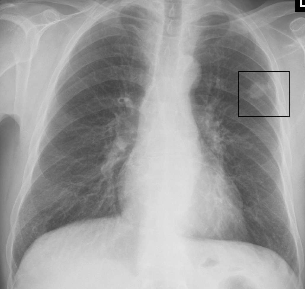
 Pulmonary Nodules Cancer Therapy Advisor
Histoplasmosis causes flu-like symptoms including fever and cough.
Small lung nodules causes. They tend to be around 019 to 098 inches 5 to 25 mm wide. Lung infections are the most common cause of lung nodules as they lead to the growth of lung tissue and inflammation. Histoplasmosis an infection that often causes lung nodules is caused by breathing in spores of a fungus often found in bird and bat droppings.
Noncancerous lung nodules can have a variety of causes including. Pulmonary nodules are common and as the spatial resolution of CT scanners has increased detection of smaller and smaller nodules has occurred which are more often an incidental finding. Pulmonary nodules are small rounded opacities within the pulmonary interstitium.
Many different lung infections such as Mycobacterium tuberculosis aspergillosis histoplasmosis coccidiomycosis and cryptococcosis can cause lung nodules to form or can result in scar tissue that forms into a nodule. These can be caused by a variety of things including. Lung nodules can be either benign non-cancerous or malignant cancer.
Certain problems such as rheumatoid arthritis can cause inflammation that is noninfectious but still causes inflammation in the lungs. A lung nodule or mass is a small abnormal area sometimes found during a CT scan of the chest. The nodule can be an indication of an active process or can be due to a scar tissue formation related from previous inflammation.
Some medical professionals will refer to them as lung spots. The most common causes overall include granulomas clumps of inflamed tissue due to an infection or inflammation and hamartomas benign lung tumors. Pulmonary Nodules Caused by Inflammation A common cause of benign pulmonary nodules is infections and inflammation in the lungs.
The nodules may show up on a chest X-ray as small white shadows. The result is the appearance of lung nodules. Cancer is one of the common causes of a lung nodule but it isnt the only common cause.
A solitary lung nodule can be found to be an arteriovenous malformation a hematoma or an infarction zone. In fact there are more than a few causes for lung nodules. The spores of this fungus are inhaled and thrive in the nutrient and oxygen rich environment of the lung tissue.
What is considered a smalllung nodule. Inflammation that is not caused by an infection-based inflammation may sometimes also develop in the lung. The nodule may represent an active process or be the result of scar tissue formation related to prior inflammation.
Inflammation from infections such as. Lung nodules are usually caused by scar tissue a healed infection that never made you sick or some irritant found in the air you breathe. The body fights off the infection leaving scar tissue which later shows up on an x-ray or CT scan as a lung nodule.
Nodules can also develop in internal tissues. Benign pulmonary nodules can have a wide variety of causes. There are different causes for benign pulmonary nodules.
What causes nodules in the lungs. The lung nodules may be caused by various conditions such as lung lesions infections and certain lung diseases. It may help you to know that nodules are detected in up to half of patients who undergo LDCT imaging.
Inflammation often occurs due to an infection or an autoimmune reaction which occurs when your body overreacts to its own tissues. When granuloma cells form around the area of inflammation in the lungs it can create a lung nodule. Infections like tuberculosis can cause inflammation.
Most are the result of old infections scar tissue or other causes and not cancer. It may also be caused by bronchial atresia sequestration an inhaled foreign body or pleural plaque. This form of fungus is often found in bird droppings making urban dwellers more likely to contract this type of infection.
Lung infections such as pulmonary tuberculosis which is caused by Mycobacterium tuberculosis granulomas which are small clumps of cells that grow because of inflammation. Lung nodules may also be caused by a host of fungal infections including histoplasmosis. Lung nodules can be harmless growths or a sign of lung cancer.
Inflammation in the lungs might occur due to a range of infections such as tuberculosis TB or fungal infections. The causes of benign lung tumors and nodules are poorly understood. Many are the result of inflammation in the lung as a result of an infection or disease producing inflammation in the body.
In these patients very few pulmonary nodules turn out to be cancer. But in general they often result from problems like these. Nodules are relatively common and the smaller they are the less trouble theyre likely to be.
Bacterial fungal or parasitic infections in the lungs.